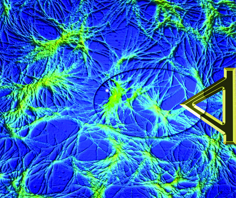
Supramolecular gels are formed by molecules that assemble to form a solid network robust enough to immobilise a volume of solvent. An exciting feature of supramolecular gels is that they respond to environmental parameters such as pH, heat, light and chemical reactions. This opens up a range of potential applications such as drug-delivery systems, tissue engineering and sensors, to name a few. The dynamic nature of these systems also presents significant challenges for characterisation. Morphological examination using typical microscopic techniques such as freeze-drying followed by electron microscopy presents obvious pathways for artifacts to be introduced. Researchers at Curtin University, in collaboration with the University of Western Australia, have developed an experimental protocol to monitor gel fibre formation on the nanoscale, dynamically and in situ (Barker E.C., Goh C.Y., Jones F., Mocerino M., Skelton B.W., Becker T., Ogden M.I. Chem. Sci. 2015, 6, 6133−8). Using an atomic force microscope tip immersed in a gelating droplet on a temperature-controlled stage, it proved possible to image supramolecular gel fibre networks in the process of forming, and disassembling, as a function of temperature. This technique provides a new approach to study the dynamics of supramolecular gel assembly and disassembly, particularly fibre−fibre interactions, at high resolution.